


Visikol HISTO-M 3D细胞球体透明化试剂


使用Visikol HISTO-M细数3D细胞球体里的细胞!
Countevery cell in your 3D model with Visikol HISTO-M
visikol HISTO-M是一种专用于三维细胞培养模型(类器官、球 状体、微组织)透明化处理的试剂盒,具有无损、快速、易 用等优点。
· 适用于厚度小于1mm的样品 · 可兼容免疫染色, · 透明化过程可逆,可继续下游分析处理
Every cell counts!
While 3D cell culture models are being adopted in the drug discoveryspace for their improved in vivo relevancy, the imaging techniquesused to characterize these models have serious room for improvement. Theproblem is due to the thickness and opacity of the 3D cell culture models. Sincethey are too thick, light cannot penetrate to the center of the tissues, and soonly the outer 2-3 layers of cells can be detected. This causes the darkcenters often seen in images of 3D cell culture models. Unfortunately, thiseffect introduces bias into results, since only the outermost cells can bedetected, and those cells are the most exposed to oxygen, nutrients, and drugcompound. Treatment with Visikol HISTO-M solves this problem.
应用案例
共聚焦成像
Clearing with Visikol HISTO-M renders the 3D model transparent, allowingfor the detection of every single cell using confocal imaging with a HighContent instrument.
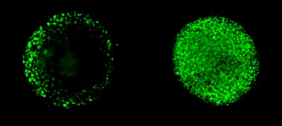
NCI-H2170 spheroids approx 250 um in diameter labeledwith nuclear stain. Left is in PBS and right is the same spheroid afterclearing with Visikol HISTO-M.
3D cellculture in PBS, not cleared. A large majority of cells are not observable.

3D cell culture after clearing withVisikol HISTO-M - every cell in the model can be detected

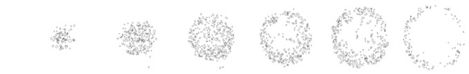
Number ofcells characterized in before and after clearing NCI-H2170 3D cell culturemodel
Resultsare clear with Visikol HISTO-M!高灵敏度,少实验误差
Significant differences between cleared and non-cleared tumor spheroidshave been observed in the dose response curves of commonantiproliferatives. The application of tissue clearing to 3D cell culturecharacterization has been shown to increase sensitivity to measure doseresponse by an order of magnitude due to the increased number of cells detectedin cleared spheroids. Furthermore, highly significant differences in doseresponse are measured between cleared and non-cleared tumor spheroids due tothe increased level of proliferation in the outermost cells.

NCI-H2170spheroids dosed with cisplatin and evaluated for cell proliferation (Ki67positive cells); A) Dose response relative to vehicle control; B) Absolute cellproliferation score
锘海生命科学成立于2017年,2020年获得国家高新技术企业资质,2021年7月被列入上海市标准化试点项目单位,项目名称为《光片照明显微镜研发与应用标准化试点》(项目编号:S21-02-025)。总部位于上海漕河泾开发区松江园区内,在北京,广州,成都,沈阳等十余座城市设有办事处, 作为“生命科学的服务者,医疗创新的推动者“,致力于打造完整的生命科学研发、制造、服务生态体系。
锘海自主研发LS18平铺光片显微镜可实现小鼠全脑、脊髓、骨骼、肾脏、肝脏、乳腺、胰腺、肺、肌肉及肿瘤等小动物完整器官3D结构呈现。“平铺光片技术”解决了传统光片显微镜中空间分辨率、光学层析能力和成像视野大小之间的矛盾,满足高通量、准确定位的荧光成像分析需求,广泛应用于脑科学、肿瘤学、药物研发、干细胞研究、组织胚胎学等各个领域。为方便广大科研工作者,我们亦提供组织透明化、免疫荧光标记、高分辨大组织3D成像、图像分析与存储,一站式科研服务。
此外,锘海还有纳米药物制备系统及纳米药物制备、检测服务—从处方筛选到制剂表征全线过程。纳米药物制备系统通过微流控芯片技术制造纳米颗粒包裹体,可包裹化药、mRNA、siRNA、DNA等小分子物质,实现该物质的体内递送,从低通量至高通量均可覆盖,适用于临床前研究和符合GMP的临床生产,并可在纳米颗粒表面添加标记物制造靶向药物。目前,锘海已服务国内多家知名药企并具备成功申报临床的案例。
我们拥有一支专业且经验丰富的研发、销售、技术和本地化服务的团队,团队中大多数人员为高学历专业硕博人才,致力于为生命科学领域的科研及企业客户提供个性化、专业化的产品、服务和整体解决方案,让生命科学更加简单、高效。
地址:上海市松江区九亭镇云凯路66号科技绿洲二期10号楼2层
电话:86-21-37827858
邮箱:info@nuohailifescience.com
网址:www.nuohailifescience.com
| 单位名称: |
|
详细地址:
上海市松江区九亭镇云凯路66号科技绿洲二期10号楼2层
|
|
qq:
2842521810
|
|
联系电话:
86-21-37827858 或 13818273779
|
| Email: |
