


长时间高分辨类器官光片显微镜


——长时间、高通量、活细胞光片成像系统
瑞士Viventis公司推出的高通量活细胞高分辨光片显微镜LS系列,是一款全新的光片成像平台,主要用于活性的光敏感样品(如卵子、胚胎、类器官等)的低光毒性、高分辨率的长期成像。
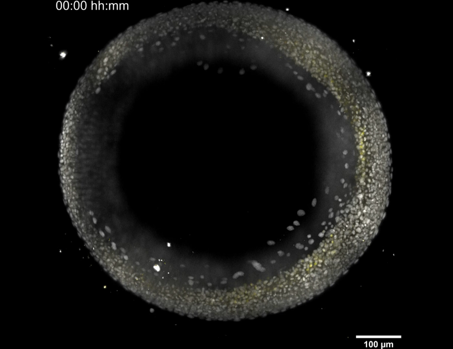
高通量活细胞高分辨光片显微镜是近些年发展起来的一种特殊的成像技术,它的照明光是一张与成像面平行的薄薄的光片,只有焦平面的样品被照亮,而光片上下的样品不受影响。该成像系统在细胞与组织层面的实时成像对于深入理解生物学行为至关重要。尤其适合对直径达300 μm的光敏样品(如卵母细胞,胚胎和类器官)进行长期实时高时空分辨率和低光毒性的观察与成像。

★ 双侧照明光片显微镜
双侧照明均可以通过软件进项控制,仅需要点击鼠标就可以控制光束的平移和旋转。光片厚度仅为1.5~6μm,且厚度可调、位置可自动校准,以适应更多的样本尺寸。配合高NA物镜,可以实现更好的穿深,更少的伪影。另外,系统配置可见激发激光器,通过检测物镜用户可对自定义样品中感兴趣的区域进行快速定位成像操作。
★ 高通量,多样品同时成像
Viventis光片显微镜可以快速对多个样品进行同时成像而无需更换样品,支持绝大多数胚胎样品并可并排摆放,方便添加培养基、加药等操作。长工作距离(样品槽尺寸>50mm),同时系统可记录多个位点并连续采集。
对于细胞球、类器官等本身较易漂浮的样本,Viventis也提供了较好的解决方案,采用了人工基底膜/水凝胶嵌入式等方案,实现上述样本的稳定成像。

★ 软件界面简洁、易于上手
Viventis系统对于光片成像初学者来说操作简单,多种模式一键切换,软件界面简洁,可以帮助您快速开启光片成像之旅,打开lightsheet大门,助力科研之路。

发表文章
2023
• Harasimov et al., Actin-driven chromosome clustering facilitates fast and complete chromosome capture in mammalian oocytes
Nature Cell Biology
• Olivetta et al., The nuclear to cytoplasmic ratio drives cellularization in the close animal relative Sphaeroforma arctica
bioRvix
2022
• Ozelci et al., Deconstructing body axis morphogenesis in zebrafish embryos using robot-assisted tissue micromanipulation.
Nature Communications
• Ishihara et al., Topological morphogenesis of neuroepithelial organoids.
Nature Physics
• de Medeiros et al., Multiscale light-sheet organoid imaging framework
Nature Communication
• Naganathan et al., Left-right symmetry of zebrafish embryos requires somite surface tension.
Nature
• So et al., Mechanism of spindle pole organization and instability in human oocytes.
Science
• Knoblochova et al., CHK1-CDC25A-CDK1 regulate cell cycle progression in early mouse embryos to protect genome integrity.
bioRvix
• Pelzer et al., Ectopic activation of the polar body extrusion pathway triggers cell fragmentation in preimplantation embryos.
bioRvix
2021
• Yang et al., Cell fate coordinates mechano-osmotic forces in intestinal crypt formation.
Nature cell Biology
• He et al., Lineage recording in human cerebral organoids
Nature Methods
• Mailand et al.,Tissue Engineering with Mechanically Induced Solid-Fluid Transitions.
Ad. Materials
• Blengini et al., Aurora kinase A is essential for meiosis in mouse oocytes.
Plos Genetics
• Rohde et al., Cell-autonomous generation of the wave pattern within the vertebrate segmentation clock
bioRvix
2020
• Rossi et al., Embryonic organoids recapitulate early heart organogenesis.
Cell Stem Cell
2019
• Serra et al., Self-organization and symmetry breaking in intestinal organoids development.
Nature
• Dumortier et al., Fracking and Ostwald ripening position the lumen of the mouse blastocyst.
Science
• Welling et al., Primed Track, high-fidelity lineage tracing in mouse pre-implantation embryos using primed conversion of photoconvertible proteins.
Elife
• Arribat et al., Mitochondria in Embryogenesis: An Organellogenesis Perspective.
Frontiers in Cell and Developmental Biology
用户单位
国内用户

典型国外用户
Quantum Design中国子公司成立于2004年,拥有一支具备强大技术背景、职业化工作作风的团队,在全权负责美国Quantum Design公司本部产品在中国销售和售后服务的同时,公司作为Quantum Design全球代理分销网的重要成员,还积极致力于发展与其他先进科学仪器制造商的合作,帮助其将产品迅速引进中国市场、发展与中国本地科学家的合作将实验方法及设备商业化。Quantum Design中国子公司的长期目标是使自身最终成为中国与世界进行先进技术及仪器交流的一个重要桥头堡。目前,Quantum Design中国子公司正立足于公司本部产品,积极致力于材料物理、纳米表征和测量技术、生物及生命科学技术领域的新业务。
Quantum Design中国 生命科学事业部 致力于引进和推介生物领域内先进设备及技术。目前已与瑞士Cytosurge公司、美国NanoView公司、波兰Novilet公司、法国Abbelight公司、法国Telight公司、德国LLS ROWIAK公司、西班牙Planelight公司、美国Delong公司等十几家生物及医药仪器设备制造商建立合作关系,帮助中国市场引进更多全球范围内的优秀设备和技术,包含但不限于新一代研发的 多功能单细胞显微操作系统、小动物自由基成像系统、全自动外泌体荧光检测分析系统、光片显微镜、3D单分子荧光成像系统、超分辨共聚焦显微系统、原位细胞3D切割成像平台、3D组织切割成像系统、低电压台式透射电子显微镜、实时无标记细胞动态分析仪等,在生命科学领域的深度探索中,助力中国科学家的项目研究和发展。
| 单位名称: |
|
详细地址:
北京市朝阳区酒仙桥路10号 恒通商务园B22座 501 室
|
|
qq:
|
|
联系电话:
010-85120280(联系我时
请说明是在生物器材网了解)
|
| Email: |
