


胶质纤维酸性蛋白单克隆抗体

基本信息

产品名称:
胶质纤维酸性蛋白单克隆抗体
英文名称:
GFAP
国产/进口:
国产
产地/品牌:
雅吉生物
型号:
33065M
参考报价:
销售商:
总点击数:
370
更新日期:
2025-09-25
产品类别:
性能参数

英文名称GFAP
中文名称胶质纤维酸性蛋白单克隆抗体
别 名Astrocyte; FLJ45472; GFAP; Glial Fibrillary Acidic Protein; Intermediate filament protein; GFAP_HUMAN.
研究领域肿瘤 细胞生物 神经生物学
抗体来源Mouse
克隆类型Monoclonal
克 隆 号7D8
交叉反应Mouse, Rat,
产品应用WB=1:500-1000 IHC-P=1:100-500 IHC-F=1:100-500 ICC=1:100-500 (石蜡切片需做抗原修复)
not yet tested in other applications.
optimal dilutions/concentrations should be determined by the end user.
分 子 量49kDa
细胞定位细胞浆
性 状Liquid
浓 度1mg/ml
免 疫 原Recombinant mouse GFAP full length:
亚 型IgG
纯化方法affinity purified by Protein G
储 存 液0.01M TBS(pH7.4) with 1% BSA, 0.03% Proclin300 and 50% Glycerol.
保存条件Shipped at 4℃. Store at -20 °C for one year. Avoid repeated freeze/thaw cycles.
PubMedPubMed
产品介绍This gene encodes one of the major intermediate filament proteins of mature astrocytes. It is used as a marker to distinguish astrocytes from other glial cells during development. Mutations in this gene cause Alexander disease, a rare disorder of astrocytes in the central nervous system. Alternative splicing results in multiple transcript variants encoding distinct isoforms. [provided by RefSeq, Oct 2008]
Function:
GFAP, a class-III intermediate filament, is a cell-specific marker that, during the development of the central nervous system, distinguishes astrocytes from other glial cells.
Subunit:
Interacts with SYNM. Isoform 3 interacts with PSEN1 (via N-terminus).
Subcellular Location:
Cytoplasm. Note=Associated with intermediate filaments.
Tissue Specificity:
Expressed in cells lacking fibronectin.
Post-translational modifications:
Phosphorylated by PKN1.
DISEASE:
Defects in GFAP are a cause of Alexander disease (ALEXD) [MIM:203450]. Alexander disease is a rare disorder of the central nervous system. It is a progressive leukoencephalopathy whose hallmark is the widespread accumulation of Rosenthal fibers which are cytoplasmic inclusions in astrocytes. The most common form affects infants and young children, and is characterized by progressive failure of central myelination, usually leading to death usually within the first decade. Infants with Alexander disease develop a leukoencephalopathy with macrocephaly, seizures, and psychomotor retardation. Patients with juvenile or adult forms typically experience ataxia, bulbar signs and spasticity, and a more slowly progressive course.
Similarity:
Belongs to the intermediate filament family.
SWISS:
P14136
Gene ID:
2670
Database links:
Entrez Gene: 281189 Cow
Entrez Gene: 2670 Human
Entrez Gene: 14580 Mouse
Entrez Gene: 24387 Rat
Omim: 137780 Human
SwissProt: Q28115 Cow
SwissProt: P14136 Human
SwissProt: P03995 Mouse
Important Note:
This product as supplied is intended for research use only, not for use in human, therapeutic or diagnostic applications.
星形胶质细胞标志物 (Astrocyte Marker)
GFAP是一个56kDa的中间丝蛋白(intermediate filament,IF),在中枢神经系统发育期是一个特异性的标志物,以区别星形细胞和其它胶质细胞。GFAP表达在皮层和海马,急、慢性皮质酮治疗时表达减少。
GFAP可以和人、大鼠、小鼠的GFAP反应,在正常和肿瘤性的星形胶质细胞阳性表达,而神经节细胞、神经元、成纤维细胞、少突胶质细胞和这些细胞来源的肿瘤细胞阴性表达,主要用于星形胶质瘤等中枢神经系统肿瘤的诊断和鉴别诊断,GFAP的缺乏可导致AD病。
| 产品图片 |
 Sample:
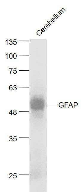
Cerebellum (Mouse) Lysate at 40 ug Primary: Anti- GFAP (bsm-33065M) at 1/1000 dilution Secondary: IRDye800CW Goat Anti-Rabbit IgG at 1/20000 dilution Predicted band size: 50 kD Observed band size: 50 kD 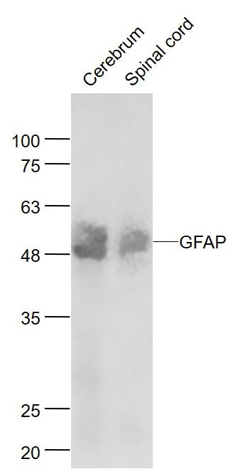 Sample:
Cerebrum (Mouse) Lysate at 40 ug Spinal cord (Mouse) Lysate at 40 ug Primary: Anti- TBX1 (bsm-33065M) at 1/1000 dilution Secondary: IRDye800CW Goat Anti-Rabbit IgG at 1/20000 dilution Predicted band size: 50 kD Observed band size: 50 kD 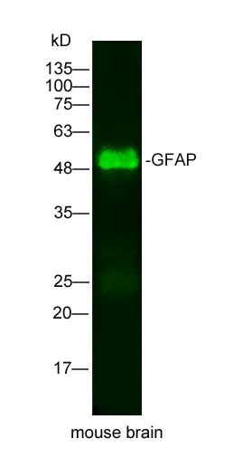 Sample: mouse brain Lysate at 25 ug
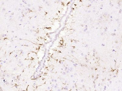
Primary: Mouse Anti-GFAP(bsm-33065M) at 1/500 dilution Secondary: IRDye800CW Goat Anti-Mouse IgG at 1/20000 dilution Predicted band size: 49kD Observed band size: 49kD 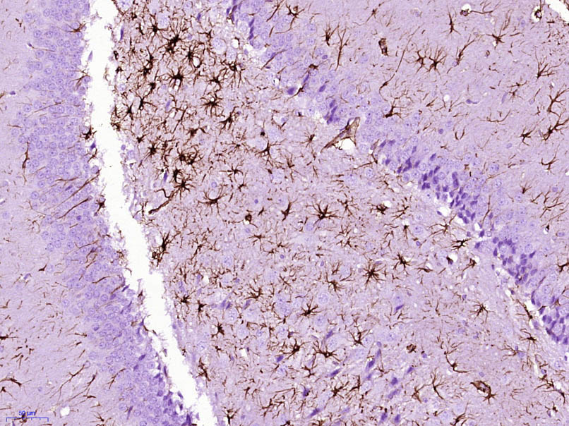 Paraformaldehyde-fixed, paraffin embedded (Rat brain); Antigen retrieval by boiling in sodium citrate buffer (pH6.0) for 15min; Block endogenous peroxidase by 3% hydrogen peroxide for 20 minutes; Blocking buffer (normal goat serum) at 37°C for 30min; Antibody incubation with (GFAP) Monoclonal Antibody, Unconjugated (bsm-33065M) at 1:400 overnight at 4°C, followed by operating according to SP Kit(Mouse) (sp-0024) instructionsand DAB staining.
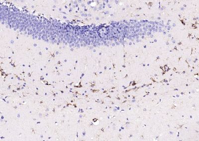
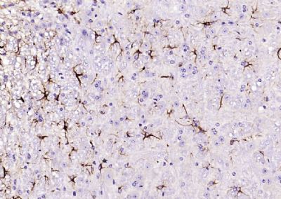 Paraformaldehyde-fixed, paraffin embedded (mouse cerebellum); Antigen retrieval by boiling in sodium citrate buffer (pH6.0) for 15min; Block endogenous peroxidase by 3% hydrogen peroxide for 20 minutes; Blocking buffer (normal goat serum) at 37°C for 30min; Antibody incubation with (GFAP) Monoclonal Antibody, Unconjugated (bsm-33065M) at 1:200 overnight at 4°C, followed by operating according to SP Kit(Mouse)(sp-0024) instructionsand DAB staining.
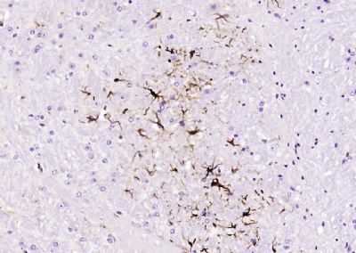 Paraformaldehyde-fixed, paraffin embedded (mouse cerebellum); Antigen retrieval by boiling in sodium citrate buffer (pH6.0) for 15min; Block endogenous peroxidase by 3% hydrogen peroxide for 20 minutes; Blocking buffer (normal goat serum) at 37°C for 30min; Antibody incubation with (GFAP) Monoclonal Antibody, Unconjugated (bsm-33065M) at 1:200 overnight at 4°C, followed by operating according to SP Kit(Mouse)(sp-0024) instructionsand DAB staining.
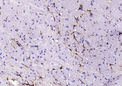 Paraformaldehyde-fixed, paraffin embedded (mouse brain); Antigen retrieval by boiling in sodium citrate buffer (pH6.0) for 15min; Block endogenous peroxidase by 3% hydrogen peroxide for 20 minutes; Blocking buffer (normal goat serum) at 37°C for 30min; Antibody incubation with (GFAP) Monoclonal Antibody, Unconjugated (bsm-33065M) at 1:200 overnight at 4°C, followed by operating according to SP Kit(Mouse)(sp-0024) instructionsand DAB staining.
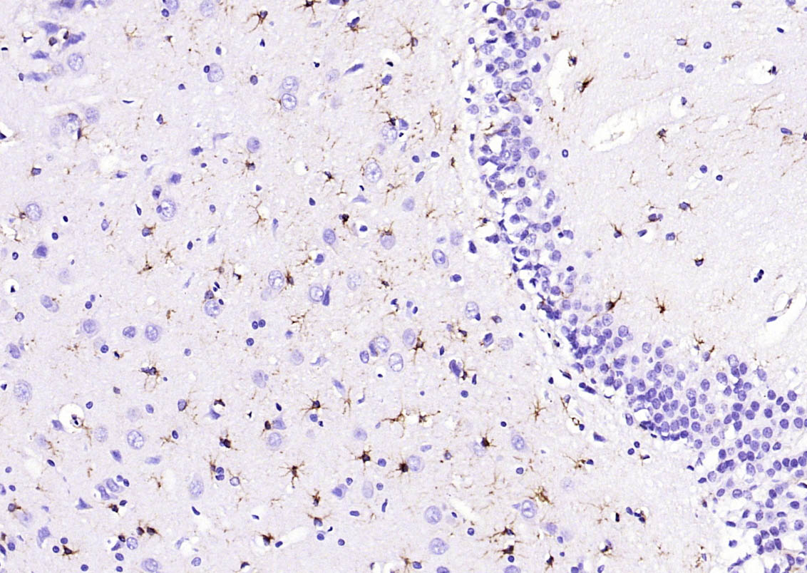 Paraformaldehyde-fixed, paraffin embedded (rat brain); Antigen retrieval by boiling in sodium citrate buffer (pH6.0) for 15min; Block endogenous peroxidase by 3% hydrogen peroxide for 20 minutes; Blocking buffer (normal goat serum) at 37°C for 30min; Antibody incubation with (GFAP) Monoclonal Antibody, Unconjugated (bsm-33065M) at 1:200 overnight at 4°C, followed by operating according to SP Kit(Mouse)(sp-0024) instructionsand DAB staining.
 Paraformaldehyde-fixed, paraffin embedded (mouse brain); Antigen retrieval by boiling in sodium citrate buffer (pH6.0) for 15min; Block endogenous peroxidase by 3% hydrogen peroxide for 20 minutes; Blocking buffer (normal goat serum) at 37°C for 30min; Antibody incubation with (GFAP) Monoclonal Antibody, Unconjugated (bsm-33065M) at 1:200 overnight at 4°C, followed by operating according to SP Kit(Mouse)(sp-0024) instructionsand DAB staining.
 Paraformaldehyde-fixed, paraffin embedded (Mouse cerebellum); Antigen retrieval by boiling in sodium citrate buffer (pH6.0) for 15min; Block endogenous peroxidase by 3% hydrogen peroxide for 20 minutes; Blocking buffer (normal goat serum) at 37°C for 30min; Antibody incubation with (GFAP) Monoclonal Antibody, Unconjugated (bsm-33065M) at 1:800 overnight at 4°C, followed by operating according to SP Kit(Mouse) (sp-0024) instructions and DAB staining.
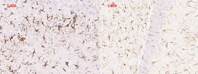 Paraformaldehyde-fixed, paraffin embedded (Rat brain); Antigen retrieval by boiling in sodium citrate buffer (pH6.0) for 15min; Block endogenous peroxidase by 3% hydrogen peroxide for 20 minutes; Blocking buffer (normal goat serum) at 37°C for 30min; Antibody incubation with (GFAP) Monoclonal Antibody, Unconjugated (bsm-33065M) at 1:400 and 1:800 overnight at 4°C, followed by operating according to SP Kit(Mouse) (sp-0024) instructions and DAB staining.
 Paraformaldehyde-fixed, paraffin embedded (Mouse brain); Antigen retrieval by boiling in sodium citrate buffer (pH6.0) for 15min; Block endogenous peroxidase by 3% hydrogen peroxide for 20 minutes; Blocking buffer (normal goat serum) at 37°C for 30min; Antibody incubation with (GFAP) Monoclonal Antibody, Unconjugated (bsm-33065M) at 1:800 overnight at 4°C, followed by operating according to SP Kit(Mouse) (sp-0024) instructions and DAB staining.
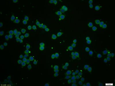 Tissue/cell: BV-2 cell; 4% Paraformaldehyde-fixed; Triton X-100 at room temperature for 20 min; Blocking buffer (normal goat serum, C-0005) at 37°C for 20 min; Antibody incubation with (GFAP) Monoclonal Antibody, Unconjugated (bsm-33065M) 1:50, 90 minutes at 37°C; followed by a conjugated Goat Anti-Mouse IgG antibody (bs-0296G-FITC) at 37°C for 90 minutes, DAPI (5ug/ml, blue, C-0033) was used to stain the cell nuclei.
|
公司简介
上海雅吉生物科技有限公司成立于2011年3月,是一家面向生命科学领域,科研机构、高校、院所及企业产品开发研究提供所需要的科研类试剂、耗材、仪器、技术服务等。包括分子生物学、免疫学、微生物学、细胞学,通过公司各个部门所有员工的共同努力在行业内拥有较高知名度,深得新老客户的厚爱,本着“优质、服务、信誉”的精神,坚持以先进的技术、优质的产品、良好的信誉,为国内外广大用户提供优质生物产品和服务。公司在重视产品质量的同时,也建立了一套集技术支持、物流、售后服务等多个部门联动服务体系,努力把我们方便、快捷、周到的服务提供给每一个客户本研究所郑重承诺:质量保证、供货及时、服务周到,公司总投资超过2000万元,并先后引进了自动化设备,组建了专业的研发技术团队。凭借着洁净化的生产车间、标准化的质量管理系统,为生产高质量的产品提供了有效保证。
雅吉生物自主研发的ELISA试剂盒,在国内众多重点实验室广泛使用,深受广大科研人员的好评,先后在权威杂志文章中被引用。
上海雅吉生物科技有限公司以极强的职业精神,积极、负责、坦诚的服务令我们的客户满意!
客户服务承诺
以“服务、客户、技术”为根本。客户是我们的核心,坚持以高质量的产品服务客户, 秉持诚信原则,重视客户的意见与感受;我们愿意为大家提供良好的售后服务。希望老师们对我们的服务及工作提出意见,您的满意是我们工作的动力,祝广大师生客户生活工作愉快!
更多产品详情可查询官网:www.yajimall.com
雅吉生物自主研发的ELISA试剂盒,在国内众多重点实验室广泛使用,深受广大科研人员的好评,先后在权威杂志文章中被引用。
上海雅吉生物科技有限公司以极强的职业精神,积极、负责、坦诚的服务令我们的客户满意!
客户服务承诺
以“服务、客户、技术”为根本。客户是我们的核心,坚持以高质量的产品服务客户, 秉持诚信原则,重视客户的意见与感受;我们愿意为大家提供良好的售后服务。希望老师们对我们的服务及工作提出意见,您的满意是我们工作的动力,祝广大师生客户生活工作愉快!
更多产品详情可查询官网:www.yajimall.com
售后服务
试剂盒售后:本公司出售的试剂盒均保质保量,质量问题均可免费退换,另提供免费代测服务。
细胞售后:
1. 细胞运输丢失、瓶身破损、培养液严重漏液等,重发;
3. 细胞收到当天以及第2,3天请拍照,未告知的视为产品合格。4-10天内出现问题,请提供细胞照片和细胞出现问题的照片以及细胞相关操作的详细步骤,并跟我公司人员及时沟通判定是否重发,具体可以参照我官网或者随货说明书相关售后条款。
相关视频
暂无
资料下载
暂无
联系方式
| 单位名称: |
|
详细地址:
上海市闵行区元江路5500号第1幢5658室
|
|
qq:
58268971
|
|
联系电话:
021-34661276
|
| Email: |
