

利用X射线显微断层扫描技术对蜥蜴脑部进行虚拟组织学研究操作流程
2024-10-16 来源:本站 点击次数:1345
Zhou, Tunhe, Yulia Dragunova, and Zegni Triki. “Brain Virtual Histology of a Lizard Species (Podarcis Bocagei) Using X-Ray Micro-Tomography and Deep-Learning Segmentation.” bioRxiv, July 6, 2024. https://doi.org/10.1101/2024.07.05.602071.
本文旨在展示如何利用X射线显微断层扫描技术(microCT)和深度学习分割,对蜥蜴(Podarcis bocagei)的脑部进行虚拟组织学研究,并提供一个详细的操作流程。
01 研究背景
02 Dragonfly软件的使用
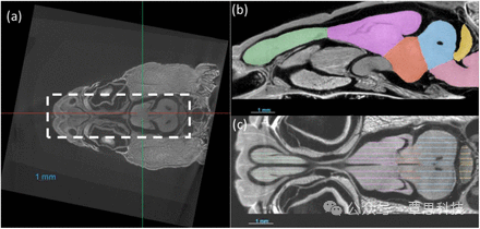 (a) The images are aligned and cropped to keep the brain for fast segmentation. (b,c) Demonstration of manual segmentation.
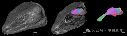
3D rendering of the X-ray microCT images showing the external and internal of the head, and the main parts of the brain. The brain regions are: the olfactory bulb (green), telencephalon (purple), diencephalon (red), midbrain (blue), cerebellum (yellow) and brainstem (pink).
03 研究方法
1. 样本准备: 对蜥蜴头部样本进行磷钨酸(PTA)染色,增强组织对比度。
2. X射线microCT扫描: 使用蔡司Xradia Versa 520扫描仪对样本进行扫描,获取高分辨率的3D图像。
3. 半手动分割训练集: 在Dragonfly中手动分割部分切片,然后使用Biomedisa软件进行随机游走插值,最后手动检查和修正。
4. 深度学习分割: 比较了Biomedisa和AIMOS两种深度学习算法,使用训练好的模型对剩余数据进行自动分割。
5. 分割评估: 使用平均dice score(DSC)和脑区体积平均相对误差(RE)评估分割精度。
04研究结果
结论
Dragonfly软件在本研究中主要用于图像预处理(对齐和裁剪)、手动分割训练集的创建,以及3D模型重建和可视化等方面,为后续的深度学习分割和分析提供了基础。
本文旨在展示如何利用X射线显微断层扫描技术(microCT)和深度学习分割,对蜥蜴(Podarcis bocagei)的脑部进行虚拟组织学研究,并提供一个详细的操作流程。
01 研究背景
- 传统的组织学方法耗时且对组织具有破坏性,而microCT可以提供高分辨率的3D图像,实现任意方向的虚拟组织学切片,减少了对样本的破坏和所需动物数量。
- 虽然磁共振成像(MRI)也被用于脑部研究,但microCT在骨骼和肌肉组织方面具有更高的对比度和分辨率,能提供更全面的头部解剖细节。
- 深度学习分割在医学影像领域发展迅速,但野生动物研究通常样本量较小。本研究旨在证明深度学习分割在小样本量下的有效性。
02 Dragonfly软件的使用
- 图像对齐与裁剪: 由于样本放置方向不一致,作者使用Dragonfly软件对图像进行对齐和裁剪,确保脑部区域一致,方便后续分析和深度学习训练。
- 手动分割训练集: 作者在Dragonfly中手动分割了约20-30个切片,作为深度学习模型的训练集。
- 3D模型重建与可视化: 作者使用Dragonfly将microCT图像重建为3D模型,并对分割后的脑区进行可视化展示。
3D rendering of the X-ray microCT images showing the external and internal of the head, and the main parts of the brain. The brain regions are: the olfactory bulb (green), telencephalon (purple), diencephalon (red), midbrain (blue), cerebellum (yellow) and brainstem (pink).
03 研究方法
1. 样本准备: 对蜥蜴头部样本进行磷钨酸(PTA)染色,增强组织对比度。
2. X射线microCT扫描: 使用蔡司Xradia Versa 520扫描仪对样本进行扫描,获取高分辨率的3D图像。
3. 半手动分割训练集: 在Dragonfly中手动分割部分切片,然后使用Biomedisa软件进行随机游走插值,最后手动检查和修正。
4. 深度学习分割: 比较了Biomedisa和AIMOS两种深度学习算法,使用训练好的模型对剩余数据进行自动分割。
5. 分割评估: 使用平均dice score(DSC)和脑区体积平均相对误差(RE)评估分割精度。
04研究结果
- 成功分割出29个样本的六个主要脑区:嗅球、端脑、间脑、中脑、小脑和脑干。
- 两种深度学习算法均只需5个训练集即可达到较高的分割精度(DSC>0.94),且与手动分割的体积测量差异约为4%。
- 不同切片方向(冠状面、矢状面和水平面)对分割精度无显著影响。
结论
- 本研究提供了一种高效、准确的蜥蜴脑部3D成像和体积测量方法。
- microCT结合深度学习分割,特别适用于样本量较小的野生动物研究。
- 该方法在生态学、进化生物学等领域具有广泛应用前景,并益于对头部其他解剖细节感兴趣的研究,以及计算机科学家开发更高效的算法。
Dragonfly软件在本研究中主要用于图像预处理(对齐和裁剪)、手动分割训练集的创建,以及3D模型重建和可视化等方面,为后续的深度学习分割和分析提供了基础。
相关文章
更多 >