

CytoViva无标记纳米高光谱显微成像系统在脂质体和外泌体领域应用介绍
2022-06-28 来源:知乎 点击次数:1895
Label-Free Liposome & ExosomeHyperspectral Microscopy
纳米尺度囊泡(如脂质体和外泌体)作为药物传递载体的研究取得了显著进展,使其在FDA的临床试验数量不断增加。大多数早期临床试验工作都集中在携带药物或基因治疗负载的工程脂质体上。然而,天然外泌体载体正在迅速取得进展。
Research of nano-scale vesicles such as liposomes and exosomes as drug delivery vectors has progressed significantly leading to increasing numbers of FDA clinical trials. Most of these early clinical trial efforts have focused on engineered liposomes carrying a drug or gene therapy load. However, rapid progress is now being made with natural exosomal vectors.
关键挑战仍然是了解这些纳米级载体靶向肿瘤细胞的功效,这通常通过内吞事件或直接细胞膜融合发生,最终将药物释放到细胞中。由于囊泡的荧光标记会干扰其任务,因此需要一种无标记的体外成像方法来确认各种囊泡-药物负载组合的这种功效。成像方法还应有助于确定囊泡样品中的适当药物负荷,并确认其与活细胞的相互作用和摄取。
Key challenges remain in understanding the efficacy of tumor cell targeting with these nanoscale carriers, which most often happens through endocytic events or direct cell membrane fusion for eventual release of the drugs to the cell. Since fluorescent labeling of the vesicle can interfere with its task, a label-free, in-vitro imaging method is required to confirm this efficacy with a variety of vesicle-drug load combinations. The imaging method should also help
determine proper drug load across the vesicle sample and confirm its interaction and uptake with live cells.
CytoViva 的增强型暗场高光谱显微镜在帮助研究人员了解囊泡载体靶向细胞以及装载药物的定时释放方面非常有效。使用这种无标签成像技术,可以:
CytoViva's Enhanced Darkfield Hyperspectral Microscope is proving to be highly effective in helping researchers understand both the targeting of vesicle carriers to cells as well as the timed release of their drug cargo. With this label free imaging technique it is possible to:
• 在体外和溶液中成像<100 nm 的无标记外泌体或脂质体
• 区分具有不同有效载荷的囊泡和空囊泡
• 使用光谱图对囊泡药物构建体进行无标记细胞运输,以识别与活细胞或固定细胞的相互作用
• Image label-free exosomes and liposomes < 100 nm in-vitro and in solution• Distinguish vesicles with differing payloads as well as empty vesicles• Conduct label-free cell trafficking of the vesicle-drug construct with spectral mapping to identify interaction with live or fixed cells
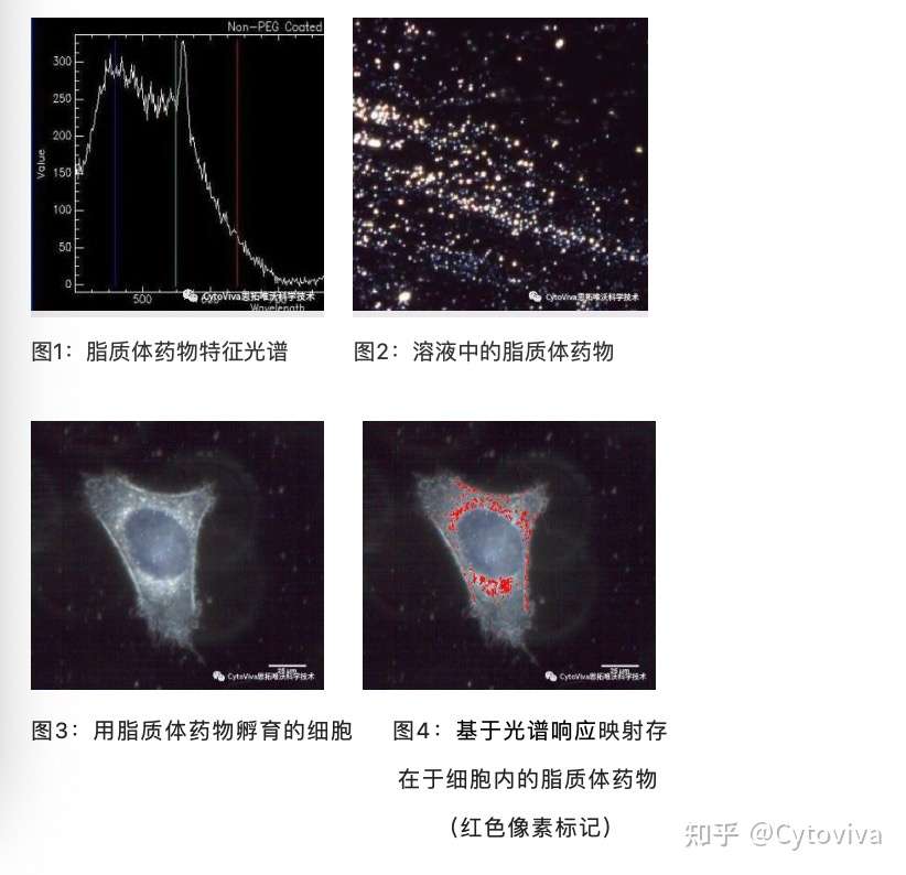
上面显示了用于此应用的 CytoViva 系统的具体示例。在此示例中,将前列腺癌特异性肽添加到负载多柔比星的脂质体中。借助 CytoViva 增强型暗场高光谱显微镜系统,研究人员能够创建负载阿霉素的脂质体(图 2)独有的参考光谱库(图 1)。请注意,图 1 中所示的参考光谱在 575nm 处有一个非常明显的峰,这与阿霉素的光谱特性一致。图 3 显示了已与脂质体构建体一起孵育的无标记肿瘤细胞。图 4 中的红色像素展示了参考光谱的光谱映射,确认了脂质体构建体在肿瘤细胞内的存在和位置。
A specific example of the CytoViva system being used for this application is shown above. In this example, a prostate cancer-specific peptide is added to a doxorubicin-loaded liposome. With the CytoViva Enhanced Darkfield Hyperspectral Microscope system, the researcher is able to create a reference spectral library (figure 1) unique to the doxorubicin-loaded liposome (figure 2). Note that the reference spectra illustrated in figure 1 has a very distinct peak at ~575nm that is consistent with the spectral properties of doxorubicin. Figure 3 shows a label-free tumor cell that has been incubated with the liposome construct. The red pixels in figure 4 demonstrate the spectral mapping of the reference spectrum confirming the presence and location of the liposomal construct within the tumor cell.
CytoViva 系统的一个主要优点是它不需要荧光标记或改变细胞及脂质体的结构即可有效的成像和分析。此外,这些样本可以使用 SynVivo for CytoViva 活细胞微流控系统进行活细胞成像。使用该系统,纳米药物递送结构可以添加到活细胞培养物中,并且可以随着时间的推移捕获图像,记录动态相互作用。该系统还可用作“模拟体内”系统,用于观察生物环境中的纳米粒子命运。
A key benefit of the CytoViva system is that it does not require fluorescent labeling or other alteration of the cell structure or the liposomes for effective imaging and analysis. Additionally, these samples can be imaged as live cells using the SynVivo for CytoViva live-cell microfluidics chamber. With this system, the nano-drug delivery construct can be added to the live cell culture and images can be captured over time, recording the dynamic interaction. This system can also be used as a "simulated in-vivo" system for observing nanoparticle fate in the biological environment.
CytoViva是由美国Auburn大学与Aetos技术有限公司合作成立,具有高校和军事公司背景,CytoViva纳米高光谱成像技术最初是由美国国防部和美国宇航局空间卫星航空成像开发的技术发展而来,该公司创造性的将该技术与增强型暗场技术结合并应用于微观层面,使其成为一个专有、集成的系统,能够在纳米尺度上对材料、药物、生命单元、活性大分子、环境污染物等进行高光谱成像及定性定量分析。
CytoViva纳米高光谱成像技术2005年一经面市,就在2006年和2007年连续两届获得著名的R&D100大奖,07年同年获得Nano50TM奖,在09年获得了两项美国专利,专利号7542203和7564623,并迅速得到全球各个国家重点实验室、科研机构及大型制药企业的认可,包括FDA, NASA, NIST, NIH, EPA, USDA, NIOSH, Lawrence Berkeley Labs, Dow Chemical,Merck, Johnson& Johnson, Stanford, Duke, Harvard等等。