

CytoViva无标记纳米高光谱显微成像系统在纳米载体开发研究中的应用
2023-02-13 来源:本站 点击次数:1950
Lipid Nano Carriers, COVID-19 and
Hyperspectral Microscopy
十多年来,纳米药物递送已经慢慢地从研究实验室发展到商业市场,并获得了少数上市批准。在2017年至2019年期间,FDA批准的纳米药物载体只有三种,然而目前有大量且不断增长的纳米药物递送应用正在获得监管部门的批准。
For over a decade, nano-drug delivery has slowly evolved out of the research laboratory and into the commercial market with a small number of regulatory approvals. Between 2017 and 2019 there were only three FDA approved nano-drug delivery constructs.1 However, there is now a large and ever-growing number of nano-drug delivery applications in the regulatory approval pipeline.
目前最引人注目的是Moderna研发的COVID-19疫苗。该疫苗使用脂质纳米颗粒作为其mRNA构建的载体,已提交FDA立即批准。预计这种纳米颗粒递送的mRNA疫苗将很快影响全球数亿人的生活。因此,这一发展有望为纳米药物递送市场提供推动作用,证明纳米材料可以安全有效地作为药物递送载体来使用。
None of these is more high profile than the Moderna COVID-19 vaccine. This vaccine, which has been submitted for immediate approval by the FDA, utilizes lipid nanoparticles as the vector for its mRNA construct. It is expected that this nanoparticle delivered mRNA vaccine will soon impact the lives of hundreds of millions of people world-wide. As such, this development is expected to provide a much needed boost to the nano-drug delivery market, proving that nanoparticles can be utilized at scale as a drug delivery vector in a safe and effective manner.
脂质纳米颗粒疗法(如Moderna疫苗)的开发需要能够验证药物疗法或脂质内及表面上纳米元素的正确摄取。此外,表征这些脂质载体如何与细胞和组织相互作用也很重要。CytoViva的增强型暗场高光谱显微镜可以成为这两项任务的有效工具。
Development of lipid nanoparticle therapies such as the Moderna vaccine requires the ability to validate proper uptake of drug therapies or other nanoscale elements within, or onto, the lipids. Additionally, it is important to demonstrate how these lipid carriers interact with cells and tissue. CytoViva's Enhanced Darkfield Hyperspectral Microscopy can be an effective tool for both of these tasks.
 |
 |
| 图1:负载AuNP的双层脂质体 | 图2:脂质体面积(黄色)和带有AuNPs的脂质体双层面积(绿色)的光谱响应 |
 |
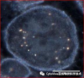 |
| 图3:在脂质体双层中映射AuNPs(红色) | 图4:巨噬细胞内LDL包裹的AuNPs |
 |
 |
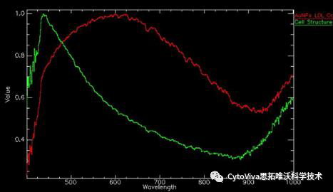
| 图5:LDL包裹的AuNPs(红色)和细胞结构(绿色)的光谱响应 | 图6:LDL包裹的AuNPs的映射(红色) |
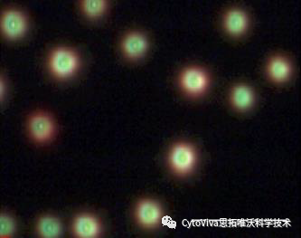
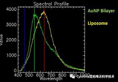
上图1说明了双层中负载AuNPs(金纳米粒子)的脂质体的增强暗场高光谱图像。金纳米粒子在它们存在于脂质体中的区域引起光谱响应的变化。这种光谱偏移如图2所示,脂质体双层中AuNPs的高光谱映射如图3(红色)所示。
Figure 1 above illustrates an enhanced darkfield hyperspectral image of liposomes loaded with AuNPs (gold nanoparticles) in the bilayer. The AuNPs cause a shift in the spectral response in areas where they are present in the liposome. This spectral shift is illustrated in Figure 2, with the hyperspectral mapping of the AuNPs in the liposome bilayer illustrated in Figure 3 (in red).
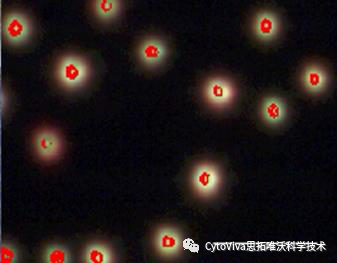
图4说明巨噬细胞中LDL包裹的金纳米粒子的增强暗视野高光谱图像。LDL包裹的金纳米粒子相对于细胞结构的独特光谱响应见图5。LDL包裹的金纳米粒子的高光谱映射如图6(红色部分)所示。
Figure 4 above illustrates an enhanced darkfield hyperspectral image of LDL encapsulated AuNPs in macrophage cells. The unique spectral response of the LDL encapsulated AuNPs versus the cell structure is illustrated in Figure 5. The hyperspectral mapping of the LDL encapsulated AuNPs is illustrated in Figure 6 (in red).
观察和光谱确认脂质内的纳米级物质或这些脂质结构被细胞摄取的能力对于有效开发和应用这些疗法至关重要。CytoViva的增强暗场高光谱显微镜已被证明是支持这些应用开发的有效方法。
The ability to observe and spectrally confirm the nanoscale cargo within the lipid or the uptake of these lipid constructs into cells is critical for effective development and deployment of these therapies. CytoViva's Enhanced Darkfield Hyperspectral Microscopy is proven to be an effective method for supporting these applications.
CytoViva是由美国Auburn大学与Aetos技术有限公司合作成立,具有高校和军事公司背景,CytoViva纳米高光谱成像技术最初是由美国国防部和美国宇航局空间卫星航空成像开发的技术发展而来,该公司创造性的将该技术与增强型暗场技术结合并应用于微观层面,使其成为一个专有、集成的系统,能够在纳米尺度上对材料、药物、生命单元、活性大分子、环境污染物等进行高光谱成像及定性定量分析。
欢迎您随时拨打公司联系电话,我们将竭诚为您提供服务!
思拓唯沃(中国)有限公司
400全国免费服务热线:400-068-0516
TEL:(010)87706572
Http://www.cytoviva.com.cn
公司地址:北京市朝阳区东三环南磨房路37号1010室